There are many types of birth defects that a baby can experience. One of them is craniosynostosis. This condition affects the shape of the skull, even the baby’s brain.
According to the Johns Hopkins Medicine page , craniosynostosis is common and occurs in one in 2,200 live births. What are the factors that can cause this birth defect ? Then, how to diagnose it? Check out the following review!
Definition
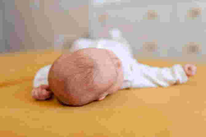
Craniosynostosis is a birth defect in which the bones in a baby’s skull fuse together too early. This occurs before the baby’s brain is fully formed. As the baby’s brain grows, the skull can become more deformed.
The spaces between the bones of a typical baby’s skull are filled with a flexible material called sutures. These sutures allow the skull to grow as the baby’s brain grows. Around age two, a child’s skull bones begin to fuse together as the sutures become bone. When this happens, the sutures are said to “close.” In a baby with craniosynostosis, one or more sutures close too early. This can limit or slow the baby’s brain growth .
When the sutures close and the skull bones fuse together too soon, the baby’s head stops growing only in that part of the skull. The rest of the skull where the sutures have not yet fused continues to grow. When this happens, the skull will have an abnormal shape, even though the brain inside the skull has grown to its normal size. Sometimes, more than one suture closes too early. In this case, the brain may not have enough room to grow to its normal size. This can cause pressure to build up inside the skull.
Symptom
Signs of craniosynostosis are usually visible at birth, but become more apparent during the first few months of a baby’s life. Signs and severity depend on how many sutures are fused and when in brain development the fusion occurs.
Signs and symptoms may include:
- The skull is deformed, with the shape depending on which sutures are affected.
- Development of a prominent hard protrusion along the affected suture, with atypical changes in the shape of the head.
- One side of a child’s face may look very different from the other side.
Other, much more common signs may include:
- Full or bulging fontanel (soft spot located on top of the head)
- Drowsiness (or less alert than usual)
- Very visible scalp veins
- Increased irritability
- High pitched cry
- Bad eating
- Projectile vomiting
- Increase head circumference
- Developmental delay.
Symptoms of craniosynostosis may resemble other conditions or medical problems, so always consult your pediatrician to clarify the diagnosis.
Reason

Often the cause of craniosynostosis is unknown, but it is sometimes linked to genetic abnormalities , such as:
- Nonsyndromic craniosynostosis is the most common type of craniosynostosis. The cause is unknown, although it is thought to be a combination of genes and environmental factors. For example, things the mother was exposed to in her environment, or what the mother ate or drank, or certain medications she took during pregnancy.
- Syndromic craniosynostosis is caused by certain genetic syndromes, such as Apert syndrome, Pfeiffer syndrome or Crouzon syndrome, which can affect the development of a baby’s skull. These syndromes usually also include other physical features and health problems.
Types of Craniosynostosis
Brachycephaly
Anterior brachycephaly involves the fusion of the right or left side of the coronal suture that runs across the top of a baby’s head from ear to ear.
This is called coronal synostosis, and it causes the normal forehead and brow ridges to stop growing. The result is a flattening of the forehead and brow ridges on the affected side, with the forehead tending to protrude too much on the opposite side. The eyes on the affected side may also be shaped differently, and there may be a flattening of the back of the head (occipital). When the fusion sutures pass through the back of the child’s skull, the result isposterior plagiocephaly.
Trigonocephaly
Trigonocephaly is a fusion of the metopic (forehead) suture. This suture runs from the top of the head in the middle of the forehead, towards the nose. Early closure of this suture can cause a prominent prominence on the forehead. Sometimes the forehead looks quite pointed, like a triangle, with the eyes set close together (hypotelorism).
Scaphoid
Scaphocephaly is the early closure or fusion of the sagittal suture. This suture runs from front to back, down the center of the top of the head. This fusion results in a long, narrow skull. The skull is long from front to back and narrow from ear to ear.
Types of Craniosynostosis Based on the Sutures that Intersect
There are also different types of craniosynostosis depending on which sutures fuse together first. Most involve fusion of a single cranial suture.
Sagittal synostosis
The sagittal suture runs along the top of the head, from the baby’s soft spot near the front of the head to the back of the head. If this suture closes too early, the baby’s head will grow long and narrow (scaphocephaly). This is the most common type of craniosynostosis.
Coronal synostosis
The right and left coronal sutures run from each ear to the sagittal suture at the top of the head. When one of these sutures closes too early, the baby may have a flat forehead on the side of the skull that closed early (anterior plagiocephaly). The baby’s eye socket on that side may also be raised and the nose may be pulled to that side. This is the second most common type of craniosynostosis.
Bicoronal synostosis
This type of craniosynostosis occurs when the coronal sutures on either side of a baby’s head close too early. In this case, the baby’s head will grow wide and short (brachycephaly).
Lambdoid synostosis
The lambdoid suture runs along the back of the head. If this suture closes too early, the baby’s head may be flattened at the back (posterior plagiocephaly). This is one of the rarest types of craniosynostosis.
Metopic synostosis
The metopic suture runs from the baby’s nose to the sagittal suture at the top of the head. If this suture closes too early, the top of the baby’s head may appear triangular, meaning narrow at the front and wide at the back (trigonocephaly). This is one of the rarest types of craniosynostosis.
Diagnosis
Craniosynostosis may be congenital (present at birth) or observed later, often during a physical examination in the first year of life. Diagnosis involves a thorough physical examination and diagnostic testing. Your child’s doctor will begin with a complete prenatal and birth history, asking about any family history of this condition or other head or facial abnormalities.
The doctor may also ask about developmental milestones, as this condition can be associated with other neuromuscular disorders. Developmental delays may require further medical follow-up for underlying problems.
During the examination, the doctor will measure the circumference of the child’s head to identify normal and abnormal ranges. Craniosynostosis can be diagnosed with a physical examination. If needed, a neurosurgeon may recommend imaging tests.
Risk Factors
The Centers for Disease Control and Prevention (CDC) reports important findings from research studies on several factors that increase the chances of having a baby with craniosynostosis:
Maternal Thyroid Disease
Mothers with thyroid disease or who are treated for thyroid disease while they are pregnant have a higher chance of having a baby with this condition, compared with mothers who do not have thyroid disease.
Certain Medications
Mothers who reported usingclomiphene citrate(fertility drugs) before or early in pregnancy are more likely to have a baby with craniosynostosis, compared with mothers who do not use these drugs.
Handling
Treating craniosynostosis involves surgery to correct the shape of the head and allow for brain growth. Early diagnosis and treatment allows the baby’s brain enough room to grow and develop.
Some treatments that can be given are:
1. Surgical Planning
Imaging studies can help surgeons develop a surgical plan. Virtual surgical planning for craniosynostosis treatment uses high-definition 3D CT scans and MRI scans of the infant’s skull to create a computer-simulated, individualized surgical plan. Based on the virtual surgical plan, a customized site is created to guide the procedure.
2. Operation
A team consisting of a head and face surgeon (craniofacial surgeon) and a brain surgeon (neurosurgeon) typically performs this procedure. The surgery can be performed endoscopically or openly. Both types of procedures generally produce excellent cosmetic results with a low risk of complications.
- Endoscopic surgery . This minimally invasive surgery may be considered for infants as young as 6 months. The surgery is performed earlier. Using a lighted tube and camera (endoscope) inserted through a small cut in the scalp (incision), the surgeon removes the affected stitches to allow the baby’s brain to grow properly. Compared with the open procedure, endoscopic surgery has smaller incisions, usually requires only one night in the hospital and usually does not require blood transfusions.
- Open surgery . Open surgery is usually done for babies older than 6 months. The surgeon makes an incision in the scalp and skull bone, then reshapes the affected part of the skull. The skull is held in place with absorbable plates and screws. Open surgery usually involves a hospital stay of three or four days, and blood transfusions are usually needed. This is usually a one-time procedure, but in complex cases, multiple open surgeries are often needed to correct the shape of the baby’s head.
3. Helmet Therapy
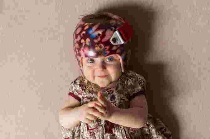
After minimally invasive surgery, office visits at regular intervals are required to fit a series of helmets to help shape the baby’s skull. The surgeon will determine the length of helmet therapy based on how quickly the shape responds to treatment. If open surgery is performed, a helmet is usually not needed afterward.
Prevention
There is no guaranteed way to prevent this birth defect. Prenatal genetic testing can show gene mutations that can cause craniosynostosis. A genetic counselor can help you understand your genetic risk and possible treatment options if your baby is born with the condition. The most important thing is for you to have regular prenatal checkups.
Although nerve damage can occur in severe cases, most children develop as expected in thinking and reasoning abilities (cognitive development) and have good cosmetic results after surgery. Early diagnosis and treatment are key to managing craniosynostosis.
Hopefully the information above can be useful for you!
***
Republished with permission from theAsianParent Indonesia


 Together Against RSV
 Pregnancy
 Parenting
 Child
Child Feeding & Nutrition
 Education
 Lifestyle
 Events
 Holiday Hub
 Aptamil
 TAP Recommends
 Shopping
 Press Releases
 Project Sidekicks
 Community
 Advertise With Us
 Contact Us
 VIP
VIP Rewards
 VIP Parents